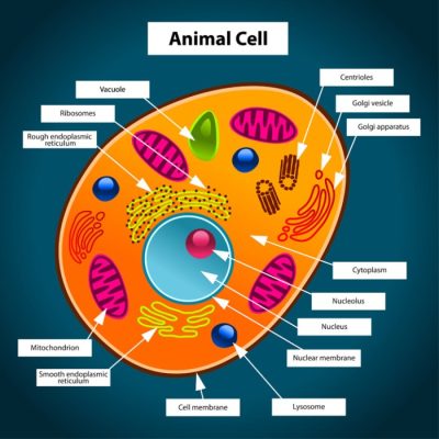
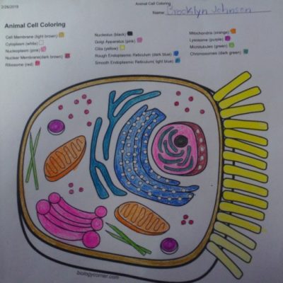
Biology Corner Animal Cell Coloring Answer Key
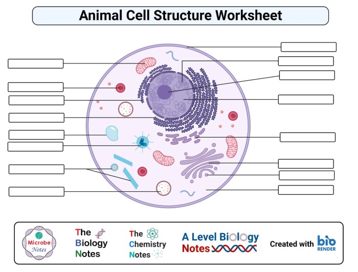
Cellular Processes Depicted in the Coloring Activity: Biology Corner Animal Cell Coloring Answer Key
Biology corner animal cell coloring answer key – This section delves into the fascinating cellular processes illustrated in the Biology Corner animal cell coloring activity. Understanding these processes is key to grasping the fundamental workings of life itself. We will explore the intricate mechanisms of cellular respiration, protein modification and transport, and the remarkable journey of protein synthesis.
Cellular Respiration in Mitochondria
Mitochondria are often called the “powerhouses” of the cell because they are the sites of cellular respiration. This process converts the chemical energy stored in glucose into a usable form of energy for the cell, ATP (adenosine triphosphate). The process occurs in several stages: glycolysis (in the cytoplasm), the Krebs cycle (in the mitochondrial matrix), and the electron transport chain (on the inner mitochondrial membrane).
Glycolysis breaks down glucose into pyruvate, which then enters the Krebs cycle. The Krebs cycle produces high-energy electron carriers (NADH and FADH2) that deliver electrons to the electron transport chain. As electrons move through the chain, a proton gradient is established across the inner mitochondrial membrane, driving ATP synthesis through chemiosmosis. Oxygen acts as the final electron acceptor, forming water.
The net result is a significant ATP yield, providing the energy needed for various cellular activities.
Golgi Apparatus: Protein Modification and Transport
The Golgi apparatus, a stack of flattened membrane-bound sacs called cisternae, plays a crucial role in modifying, sorting, and packaging proteins synthesized by ribosomes. Proteins synthesized on ribosomes attached to the endoplasmic reticulum (ER) enter the Golgi apparatus through the cis face. As proteins move through the Golgi cisternae, they undergo various modifications, such as glycosylation (addition of sugar chains) and proteolytic cleavage (removal of amino acid sequences).
These modifications are crucial for protein function and targeting. Once modified, proteins are sorted and packaged into vesicles at the trans face of the Golgi. These vesicles then transport the proteins to their final destinations, which may be other organelles within the cell, the plasma membrane, or secretion outside the cell. The Golgi apparatus acts as a central processing and distribution center for proteins, ensuring they reach their correct locations and perform their designated functions.
Protein Synthesis: From DNA to Protein Export
Protein synthesis is the process of creating proteins from the genetic information encoded in DNA. It begins in the nucleus with transcription, where the DNA sequence of a gene is copied into a messenger RNA (mRNA) molecule. The mRNA then leaves the nucleus and travels to a ribosome in the cytoplasm or on the rough endoplasmic reticulum. Translation occurs at the ribosome, where the mRNA sequence is read and used to assemble a chain of amino acids, forming a polypeptide.
The sequence of amino acids is determined by the mRNA sequence, which is ultimately dictated by the DNA sequence of the gene. Once synthesized, the polypeptide chain may undergo folding and modifications before becoming a functional protein. If the protein is destined for secretion or insertion into a membrane, it will be transported through the endoplasmic reticulum and Golgi apparatus before being packaged into vesicles and exported from the cell.
This intricate process ensures the precise synthesis and delivery of proteins essential for cellular function and overall organismal survival.
Microscopic Visualization of Animal Cells
Observing animal cells under a microscope unveils the intricate details of their structure and function. The magnification used significantly impacts the level of detail visible, and staining techniques are crucial for enhancing contrast and identifying specific organelles.
At low magnification (e.g., 4x or 10x), the overall shape and size of the cell are apparent. The cell membrane, a thin, delicate boundary, may be visible as a faint Artikel. Internal structures will be largely indistinct at this magnification. Increasing magnification to 40x reveals more cellular details. The nucleus, a large, typically round or oval structure, becomes clearly visible, often appearing as a darker, denser area within the cell.
The cytoplasm, the jelly-like substance filling the cell, can be observed as a lighter, more granular area surrounding the nucleus. However, individual organelles remain largely unresolved at this magnification. Higher magnifications (e.g., 100x using oil immersion) are necessary to resolve finer details.
Staining Techniques for Enhanced Visibility
Staining techniques are essential for improving the visibility of cellular components. Different stains bind to specific cellular structures, increasing contrast and allowing for better visualization. For instance, hematoxylin stains the nucleus a dark purple or blue, while eosin stains the cytoplasm a pinkish hue. These are commonly used in histological preparations, enabling clear differentiation between the nucleus and cytoplasm.
Other specialized stains can target specific organelles like mitochondria or the Golgi apparatus, providing more detailed information about their morphology and location within the cell. The choice of stain depends on the specific organelles of interest and the desired level of detail.
Educators seeking supplemental materials for the Biology Corner animal cell coloring worksheet might find the answer key challenging to locate. However, a fun and engaging way to reinforce learning about cell structures is through visual aids, such as those found by visiting print coloring pages animals for inspiration. This can help students visualize the shapes and relative sizes of organelles before tackling the more complex Biology Corner worksheet and its corresponding answer key.
Organelle Appearance Under the Microscope
At high magnification, various organelles become discernible. The nucleus, stained intensely, shows its characteristic nuclear membrane and potentially nucleoli, appearing as smaller, darker spots within the nucleus. Mitochondria, the “powerhouses” of the cell, appear as small, rod-shaped or oval structures scattered throughout the cytoplasm. Their appearance might vary depending on the metabolic state of the cell; active mitochondria may appear more elongated.
The endoplasmic reticulum (ER), a network of interconnected membranes, may appear as a faintly stained, somewhat reticulated structure. The Golgi apparatus, involved in protein modification and packaging, may appear as a stack of flattened sacs, often located near the nucleus. Lysosomes, involved in cellular waste disposal, are generally too small to be resolved clearly with a light microscope unless employing specialized staining techniques.
Interpreting Microscopic Images of Animal Cells
Interpreting microscopic images requires a systematic approach. First, assess the overall cell morphology: its shape, size, and any noticeable irregularities. Then, identify the major organelles: the nucleus, with its characteristically dark staining; the cytoplasm, filling the cell’s interior; and any visible organelles like mitochondria, if their staining allows for clear identification. Careful observation and comparison with known images and diagrams are essential for accurate identification.
The magnification used should always be noted, as it directly impacts the level of detail visible. For example, a clear image at 100x oil immersion would reveal far more detail than an image at 40x. Understanding the staining technique employed is also crucial, as different stains highlight different cellular components. By combining these observations, a comprehensive understanding of the cell’s structure and potential function can be derived.
Beyond the Basics
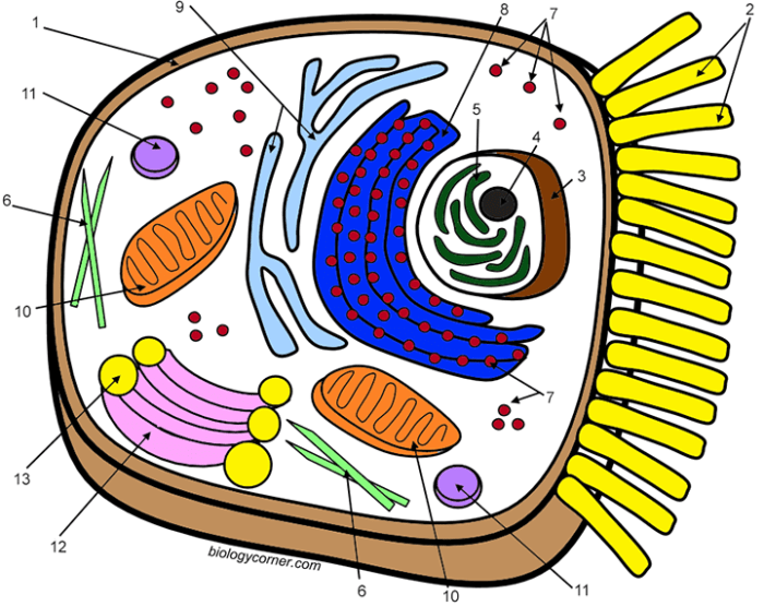
Unlocking a deeper understanding of animal cells requires exploring their intricate inner workings beyond the basic organelles. This section delves into advanced concepts crucial for comprehending cellular function and interaction within complex organisms. We will examine the dynamic cytoskeleton, the diverse array of cell junctions, and the sophisticated mechanisms of cell signaling.The cytoskeleton is a complex network of protein filaments that provides structural support, maintains cell shape, and facilitates movement.
It’s not a static structure; instead, it’s a dynamic system constantly assembling and disassembling to meet the cell’s needs. This remarkable adaptability allows cells to change shape, migrate, and divide.
The Cytoskeleton: Structure and Function
The cytoskeleton comprises three major components: microtubules, microfilaments, and intermediate filaments. Microtubules, the largest filaments, are hollow tubes made of tubulin proteins. They form the tracks along which organelles and vesicles move within the cell, contributing to intracellular transport. Microfilaments, composed of actin proteins, are involved in cell shape changes, muscle contraction, and cell division. Intermediate filaments, made of various proteins depending on the cell type, provide tensile strength and anchor organelles.
For example, the keratin filaments in skin cells contribute to their resilience. The dynamic interplay between these three filament types allows the cell to respond effectively to its environment.
Cell Junctions: Connecting Cells in Tissues
Animal cells rarely exist in isolation; they form tissues and organs through specialized cell-cell junctions. These junctions provide structural support, maintain tissue integrity, and facilitate communication between adjacent cells.There are three main types: tight junctions, adherens junctions, and gap junctions. Tight junctions seal the spaces between cells, preventing leakage of molecules between them. This is crucial in tissues like the lining of the intestines, preventing the passage of harmful substances into the bloodstream.
Adherens junctions connect cells through transmembrane proteins, providing strong adhesion between cells and contributing to tissue strength. These junctions are essential in tissues subjected to mechanical stress, such as the skin and heart muscle. Gap junctions form channels that directly connect the cytoplasm of adjacent cells, allowing the rapid exchange of small molecules and ions. This direct communication is vital for coordinated cellular activities in tissues like cardiac muscle, where synchronized contractions are essential.
Cell Signaling and Communication
Cells constantly communicate with each other and their environment through complex signaling pathways. This communication is crucial for coordinating cellular activities, regulating growth and development, and responding to external stimuli. Cell signaling involves the release of signaling molecules, such as hormones or neurotransmitters, that bind to specific receptors on the target cell’s surface or inside the cell. This binding triggers a cascade of intracellular events, ultimately leading to a cellular response.
For instance, the binding of adrenaline to receptors on heart muscle cells increases heart rate. The complexity of these pathways allows for precise control and integration of cellular responses. Disruptions in cell signaling can have serious consequences, contributing to diseases such as cancer and diabetes.
Illustrative Examples

This section provides detailed descriptions of key animal cell organelles, focusing on their structure and function. Understanding these components is crucial to comprehending the overall operation of the cell. We’ll explore the lysosome, the endoplasmic reticulum (both rough and smooth), and the nuclear envelope, highlighting their unique characteristics and contributions to cellular processes.
Lysosome Structure and Function
Lysosomes are membrane-bound organelles found within animal cells. They are spherical in shape, typically ranging from 0.1 to 1.2 µm in diameter. Their size can vary depending on the cell type and its metabolic activity. Internally, lysosomes contain a variety of hydrolytic enzymes, including proteases, lipases, and nucleases, which are responsible for breaking down various cellular components.
These enzymes function optimally in an acidic environment, maintained by a proton pump within the lysosomal membrane. The lysosome’s single membrane acts as a protective barrier, preventing the release of these potent enzymes into the cytoplasm, where they could cause significant damage. Their primary function is waste breakdown and recycling of cellular materials, including damaged organelles and ingested pathogens.
This process is essential for maintaining cellular homeostasis and preventing the accumulation of potentially harmful substances.
Endoplasmic Reticulum Structure and Function, Biology corner animal cell coloring answer key
The endoplasmic reticulum (ER) is an extensive network of interconnected membranous sacs and tubules that extends throughout the cytoplasm. It appears as a complex, labyrinthine structure under a microscope. The ER is broadly categorized into two types: rough endoplasmic reticulum (RER) and smooth endoplasmic reticulum (SER). The RER is studded with ribosomes, giving it a rough appearance. These ribosomes are responsible for protein synthesis, and the RER plays a crucial role in the folding, modification, and transport of these proteins.
In contrast, the SER lacks ribosomes and appears smooth. It is involved in lipid synthesis, carbohydrate metabolism, and detoxification of harmful substances. The interconnected nature of the ER allows for efficient transport of molecules between different cellular compartments. The RER’s association with ribosomes highlights its central role in protein synthesis and processing, while the SER’s smooth structure reflects its involvement in lipid metabolism and detoxification.
Nuclear Envelope Structure
The nuclear envelope is a double membrane structure that surrounds the nucleus, separating the genetic material from the cytoplasm. This double membrane is composed of two lipid bilayers, an inner and an outer membrane, separated by a narrow perinuclear space. The outer membrane is continuous with the endoplasmic reticulum and often studded with ribosomes. Embedded within the nuclear envelope are numerous nuclear pores, which are large protein complexes that regulate the transport of molecules between the nucleus and the cytoplasm.
These pores selectively allow the passage of specific molecules, such as mRNA and proteins, while preventing the free diffusion of others. The double membrane structure, along with the selective permeability of the nuclear pores, maintains the integrity of the nucleus and ensures the controlled exchange of materials between the nucleus and the cytoplasm. The overall appearance is that of a double-layered, porous membrane surrounding the nucleus.

